
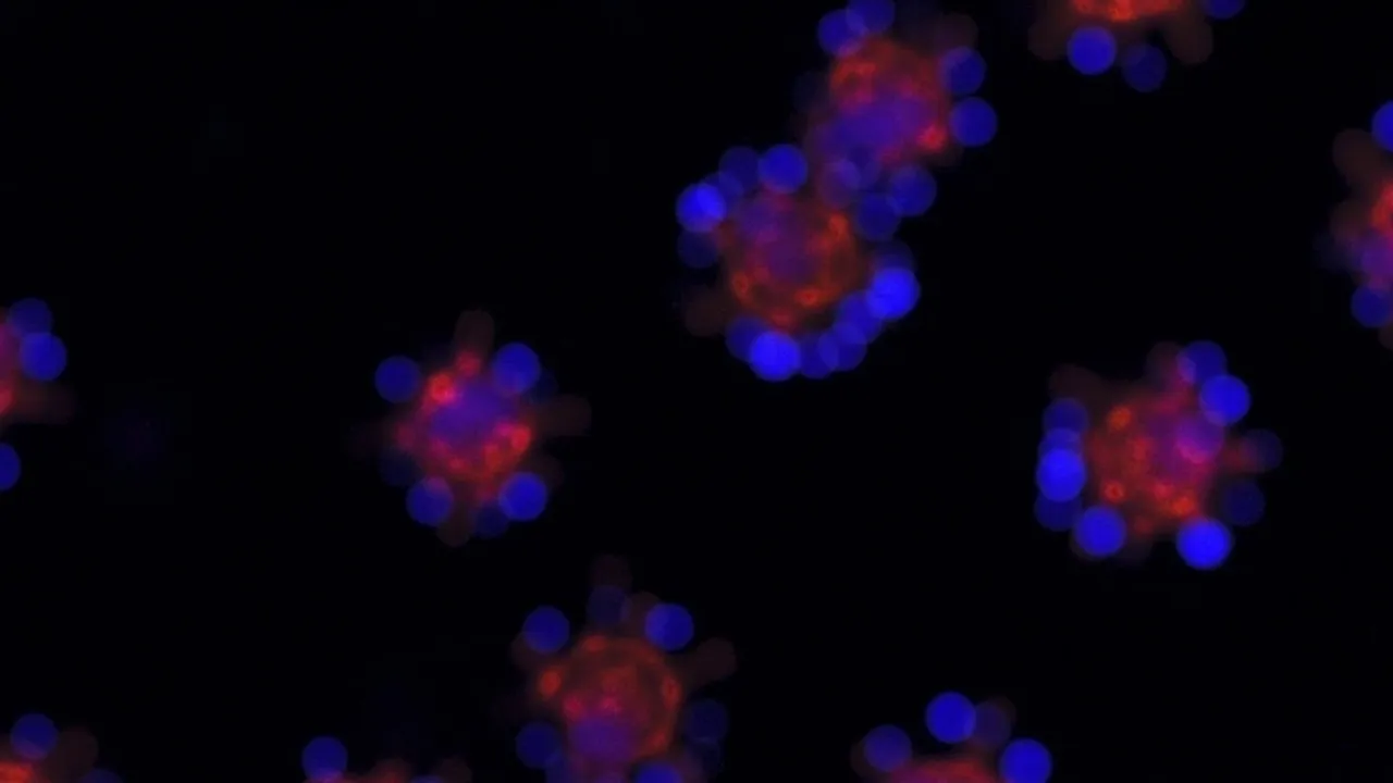
Imagine Watching a City from Inside a Bus
Okay, here's something that'll blow your mind if you love science stuff.
For decades, scientists have used powerful microscopes to zoom in on cells — those tiny biological machines that make up everything living. But here's the problem: to see anything clearly, researchers had to stain cells with fluorescent dyes. It's like putting on sunglasses so you can see better. Except the sunglasses might change how the cell behaves, and they only let you look at a few things at once.
Now, researchers at Stanford University have built something new. A microscope that watches living cells doing their thing without needing those fluorescent labels at all. And honestly? This feels like a bigger deal than it's getting credit for.
What the New Microscope Can Do
The system is called Interferometric Image Scanning Microscopy, which is a mouthful, so let's just call it iISM. What makes it special?
It can watch multiple structures inside a living cell moving, changing, and interacting all at the same time — with about 120 nanometers of resolution. For context, a nanometer is a billionth of a meter. We're talking incredibly small here.
But the real magic isn't just the resolution. It's that scientists can observe these cellular components over extended periods without frying the cell with too much light or accidentally changing how it behaves. Lower light exposure means less photodamage. Less photodamage means happier, more natural cells. More natural cells mean better science.
Why Fluorescent Labels Have Been a Headache
Let me break down why this matters so much, because I think the technical stuff can obscure how genuinely frustrating the old approach was for researchers.
Traditional fluorescence microscopy works by attaching glowing molecules to specific structures inside cells. Those molecules emit light when excited, making the target structures visible. Sounds great, right?
Except:
- You can only label a few structures at once
- The labels can fade through something called photobleaching
- Adding labels can actually change how the structures behave (oops)
- And you're only seeing labeled things, missing the broader cellular context
The Stanford team's approach avoids all of that. Think of it like switching from a flashlight that only shows you certain objects to a general light that lets you see everything in a room. You're not replacing what fluorescence does — you're complementing it with something it couldn't do before.
How Does It Actually Work? (No Engineering Degree Required)
Here's where it gets cool. The microscope uses a phenomenon you experience every single day: light scattering.
You know why the sky looks blue? As sunlight passes through Earth's atmosphere, tiny molecules scatter shorter blue wavelengths more than longer red wavelengths. That's why overhead looks blue while the sunset appears reddish — the light from the sun is being scattered all around you.
Interferometric scattering microscopy does the same thing on a teeny, tiny scale. A laser shines into a cell, and tiny structures inside scatter some of that light. The scattered signal is super weak, so they use a second laser beam to amplify it — that's the "interferometric" part.
The result? Clear images of cellular machinery going about its business without anyone having injected anything or added any labels.
What's This Mean for Science?
According to the researchers — including W.E. Moerner, who won the 2014 Nobel Prize in Chemistry for super-resolution fluorescence microscopy — this could help us:
- Study how cancer cells respond to potential drugs
- Watch how disease-causing organisms interact with cells
- Observe plant cells talking to microorganisms
- Better understand processes that were previously too difficult to study
The key phrase is "complementary." iISM isn't meant to replace fluorescence microscopy. It's meant to work alongside it. As first author Michelle Kueppers put it, "If we use the strengths of fluorescence for molecular specificity and the strength of iISM for label-free context and dynamics, we can really start tackling questions that have been difficult to address before."
To me, that sounds like the best possible outcome. Not one technology winning over another, but scientists having more tools in their toolkit to understand how life actually works at the most fundamental level.
Watching Life Unfold
There's something almost meditative about the idea of watching a living cell for hours, seeing its structures drift and interact without any artificial interruption. We're literally watching biology happen in near-real-time, unfiltered.
That feels significant. Every time we develop a new way to observe the natural world more clearly, we discover things we never expected. This microscope probably won't be the exception.
Source: ScienceDaily