The Ultimate Microscopic Showdown
Imagine if you could zoom in and watch a tiny assassin creep up on its target, make contact, and execute a perfectly calculated strike without harming anyone nearby. That's basically what your immune system's killer T cells do every single day. Now, thanks to some seriously clever microscopy work, scientists have finally captured this drama in stunning 3D detail.
The team at the University of Geneva and Lausanne University Hospital managed something researchers have been chasing for years: actually seeing how these immune cells operate inside real tumor tissue. And let me tell you, what they found is way more interesting than just confirming what we already suspected.
The Problem Nobody Could Solve (Until Now)
Here's the thing about studying killer T cells in action—it's ridiculously hard. These cells are tiny, delicate, and work in three dimensions inside even tinier human cells. Traditional ways of preparing samples for microscopy literally destroy the structure you're trying to observe. It's like trying to photograph a spider web by blasting it with a fire hose.
Scientists faced an impossible choice: get high-resolution images but lose the cell's natural structure, or keep everything intact but miss the crucial details. Neither option was good enough for the kind of breakthrough research we need.
Enter: Cryo-Expansion Microscopy (The Game Changer)
The researchers deployed a technique with a name that sounds like it belongs in a sci-fi movie: cryo-expansion microscopy, or cryo-ExM for short. Here's what makes it so clever:
Instead of using traditional sample preparation (which wrecks everything), they instantly freeze the cells at extreme speeds. This creates something called a "vitreous state," where water basically turns into glass without forming ice crystals. It's like hitting the pause button on biology itself.
Then they use a special absorbent gel to physically expand the sample, which lets them zoom in on the internal structures while keeping everything in its natural configuration. It's mind-blowing stuff—kind of like discovering you can make a LEGO set bigger without changing any of its proportions.
What They Actually Discovered
Once they could finally see what was happening, some genuinely surprising details emerged.
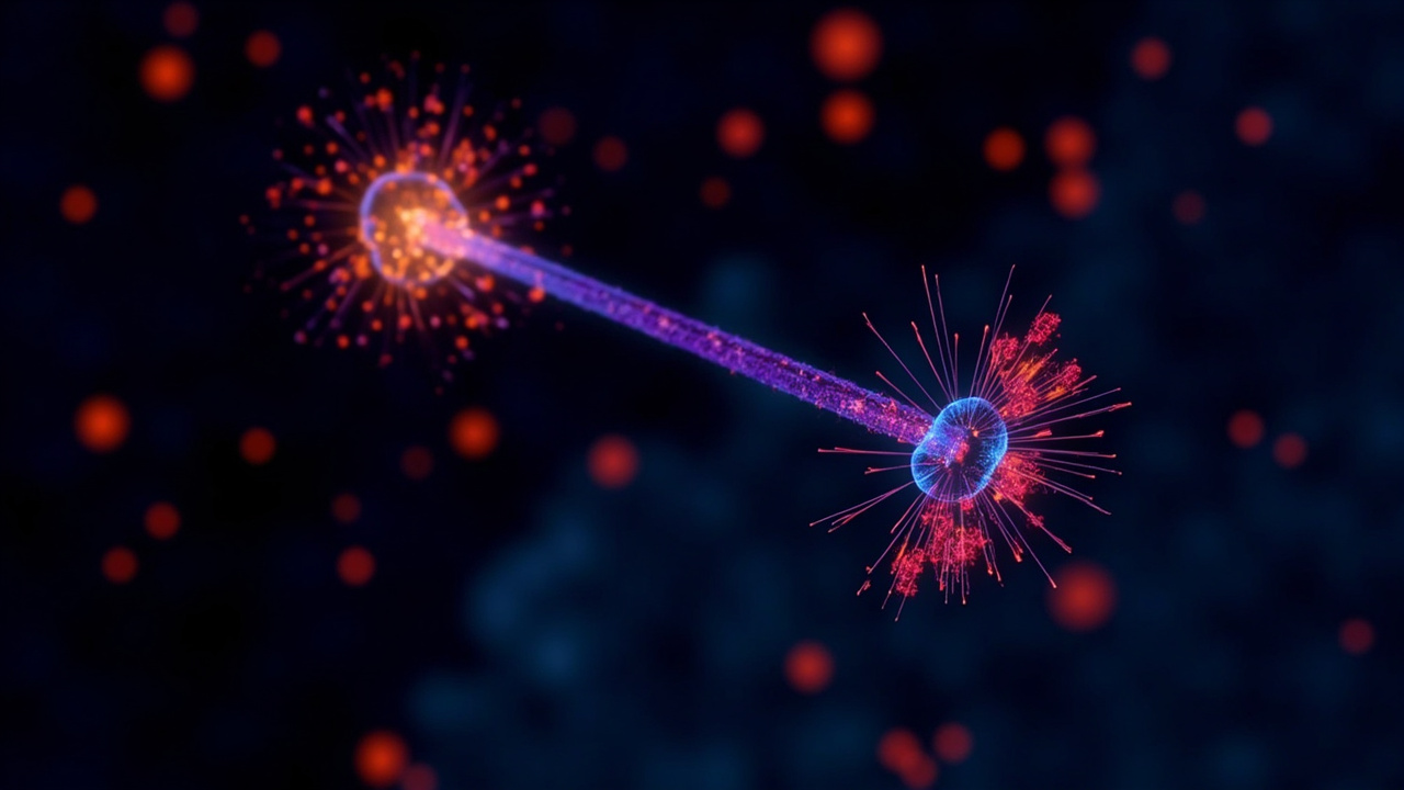
The Dome Structure: When a killer T cell makes contact with its target, the cell membrane doesn't just flatten against it—it forms a kind of dome shape. This dome's architecture seems directly connected to how the cell sticks to its target and how everything is organized inside. That's new information that could unlock why some immune attacks work better than others.
The Granule Variations: The toxic granules (basically the ammunition these cells use to kill cancer) don't all look the same under magnification. Some have one core where the killing molecules are stored, while others have multiple cores. This variation could be important for understanding why different immune responses have different effectiveness.
The Really Big Deal: Testing in Real Tumors
Here's where things get genuinely exciting. The researchers didn't just study isolated cells in petri dishes—they applied this technique directly to actual human tumor samples. This means they could watch killer T cells infiltrating real tumors and deploying their weapons in actual clinical context.
This is crucial because lab conditions and real-world biology aren't always the same. What looks effective in a controlled experiment might behave totally differently inside an actual tumor with all its complications. Now researchers have a way to study immune responses exactly where they happen.
Why This Matters for Cancer Treatment
Cancer immunotherapy has become one of the most promising approaches in modern medicine, but we still don't fully understand what makes some immune attacks succeed while others fail. By seeing exactly how killer T cells operate at the nanometer scale—inside real tumors—scientists can start figuring out:
- What structural features make a killer T cell more effective
- How tumors might be interfering with immune attacks
- How we might be able to engineer better immune responses
- Why some patients respond to immunotherapy while others don't
This isn't just academic curiosity. These insights could directly translate into better treatments, smarter immunotherapy combinations, and personalized approaches to cancer care.
The Bigger Picture
What I find most interesting about this work is that it's a perfect example of how scientific breakthroughs often come from asking "how can we actually see this thing?" rather than "what will happen if we try this treatment?" By developing better tools to observe nature, we sometimes discover things that completely change how we approach problems.
The human immune system is far more sophisticated than our current understanding gives it credit for. Every time we get a clearer view of how it works, we find new layers of complexity and opportunity. This research suggests that there's still plenty of room to improve cancer immunotherapy—we just needed to watch the process more carefully.
Pretty cool that we're at a point where scientists can literally zoom into human cells at near-atomic resolution and watch the drama of survival unfold. And even cooler that this knowledge might help us outsmart one of humanity's toughest diseases.